Warning
  • Epidural infusions for acute pain may ONLY be managed in the following areas:
    1. ITU
    2. Surgical High Dependency
    3. Recovery, Main Theatres
  • The responsibility for ensuring safe and effective epidural analgesia after surgery rests with the Anaesthetist the instituted the epidural and with the Department of Anaesthesia. Changes to the epidural prescriptions may only be authorised by an
    Anaesthetist or the Acute Pain Nurse.
  • Epidural infusions are delivered by a dedicated Epidural pump only.
  • Insertion of epidural catheter is an aseptic technique, performed in theatre. The catheter can remain in-situ for a maximum of 48 hours. If epidural analgesia is required for longer, the decision must be agreed by the anaesthetist who inserted the epidural or the On Call/Duty consultant anaesthetist.
  • All routine surgical problems can be dealt by the Surgical Medical Staff. For example: Hypotension; Nausea and Vomiting; Urine Collection; Itching
The Acute Pain Team (bleep 1003) or in their absence, the On Call/Duty anaesthetist should be contacted for any of the following epidural problems: Inadequate Pain Relief; Motor Block; Hypotension that has not improved with treatment; High sensory block; Signs of Local Anaesthetic toxicity; Patient sedation or confusion.

Setting up an epidural infusion

  • Any of the following who have received training in the use of the epidural pump: Anaesthetists; Anaesthetists assistants; Acute Pain Nurses

Procedure

  • Obtain 200ml bag of 0.125% levobupivacaine.
  • Attach the infusion bag to the epidural giving set.
  • Programme the Epidural pump and purge the line according to the manufacturer’s instruction and as per training.
  • On arrival in recovery, two recovery nurses must check the pump programme against the prescription sheet to ensure that it is correct.
  • Prior to discharge from recovery the pump and the prescription must be checked with the ward nurse

Dressings and infection control

  • Epidural catheters must be inserted using full aseptic technique and should preferably be sited in theatres or ITU.
  • The Acute Pain Service can advise on the epidural fixation device to be used.
  • The epidural filter should be secured to the upper arm or chest wall with a padded dressing, preventing the catheter from being pulled out of the Portex connector. If the line does become disconnected at this point (i.e. between the filter and the patient) the epidural catheter is inevitably contaminated and must be removed.
  • Epidural infusion bags should not be changed until they are empty. The infusion tubing should not be disconnected for any purpose other than air in the giving set.
  • The skin exit site should be inspected every nursing shift. If there are any signs of infection (tenderness, inflammation or exudation), the Acute Pain Nurse or Anaesthetist should be informed. If the epidural is removed, the tip should be sent to bacteriology for culturing.
  • The epidural catheter may remain in situ for 48 hours post-operatively. If the epidural needs to remain for any longer, this must be discussed with the anaesthetist who initially sited the epidural, or (in their absence) with a Consultant Anaesthetist.

This helps us identify which nerves have been blocked by the local anaesthetic. We assess thermo receptors at a dermatomal level using a cold stimulus (such as ice) only. It is useful to have a supply of ice cubes in the freezer compartment of your fridge. The ice cube can be placed in either a disposable glove and in a gauze for single patient use.

Nursing management

  • Children with epidural infusions may only be nursed on designated wards and be cared for by registered nurses who:
    - holds a valid current (3 yearly) certificate of attendance of the Acute Pain Service epidural lecture.
    - declares her/himself to be competent
  • If an opiate has been added to the epidural local anaesthetic infusion then no other opioids should be given whilst epidural infusion is in progress.
  • All patients who have an epidural infusion should have a patent IV cannula in case of adverse reactions
  • If epidural opiates are being used, naloxone should be readily available on the ward and registered nurses should know where it is located

Monitoring epidural infusions

  1. Pulse, Respiratory rate, Oxygen Saturations and Sedation score should be recorded on the PEWS chart:
    In recovery: every 15 mins
    On ward: 1/2 hourly for first 1 hour
    Hourly thereafter until; the epidural is discontinued
    If the child receives a “top-up” of levobupivacaine stronger that 0.125%, the patient must have their BP recorded every 5 minutes for 30 minutes.
  2. Motor score must be recorded on the epidural chart:
    In recovery: before returning to the ward
    On ward: 1/2 hourly for 2 hours
    4 hourly thereafter
  3. Sensory level must be recorded on the epidural chart:.
    In recovery: on anaesthetist’s instruction and before transfer to the ward.
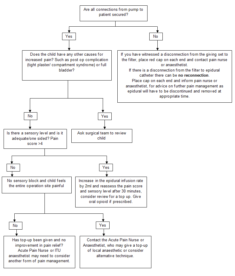
    On ward: 6hourly and if any of the following are noted:
    • inadequate pain relief
    • hypotension
    • increased motor block
    • nausea and vomiting
    • before restarting the epidural infusion if it was stopped due to high sensory level
    • one hour after any increase in the epidural infusion.
  4. Temperature should be recorded on the PEWS chart every 4 hours or more frequently depending on the child’s condition.
  5. Pump Recordings must be recorded on the epidural chart as per hospital policy, ensuring that the amount delivered and the amount remaining add up to the original starting total.
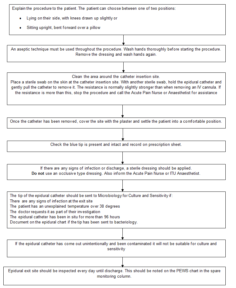
  6. Epidural Exit Site: While the epidural infusion is in progress, the exit must be checked on every shift for signs of leakage or infection. Once the epidural catheter has been removed, the exit site must be checked daily until the patient is discharged from hospital. This must be documented on the PEWS chart. Any signs of infection must be reported to the Acute Pain Team or On Call/Duty anaesthetist.
  7. Temperature must be recorded on the PEWS chart every 4 hours (or more frequently, if the patients condition requires it)
  8. Pump recordings must be recorded on the epidural chart as per Hospital policy, ensuring that the 'amount delivered' and the 'amount remaining' add up to the original starting total.

Measuring sensory levels

How to perform the sensory level measurement.

  • Explain the procedure to the child.
  • Place the ice cube on a part of the child’s body that is not affected by the epidural e.g. the patient’s forehead or hand and ask if it feels cold.
  • Next, place the ice cube below the level of the wound area and work upwards, placing the ice cube at each dermatomal level and on both sides of the body, thus checking that the block is bilateral. (This can also be done on the patients back).
  • When the child feels the ice as cold (the same as when placed on their forehead) it is usually a good indication of how far the block has spread. Using the body chart on the epidural laminated guidelines for reference, you will be able to determine the dermatomal level.
  • If the child feels the cold stimulus all over the area that is being tested and the child’s pain is controlled do not be alarmed. This occasionally occurs when weak local anaesthetic solutions are used which do not have an effect on the thermo receptors.

Measuring motor block

Epidural infusions used for post-operative pain relief should not cause the patients legs to become weak. The motor nerves are not normally affected by the weak solution of local anaesthetic used but there may be residual motor block if the patient has had a recent top-up in theatre/recovery but this should resolve within 2-4 hours. Patients in bilateral leg cast should be able to wiggle their toes without problem.

This is a side effect that must be acted upon and treated.

  • All patients must have their motor block score performed and recorded at the same time as their vital sign observations.
  • If the motor block is 2 or 3, the epidural must be temporarily stopped and the Acute Pain Nurse or the Anaesthetist contacted. 

Motor Block Score
0 = Full movement
1
= Inability to raise extended leg but able to bend knee
2
= Inability to bend knee but able to flex ankle
3 = No movement

Changing an epidural bag

Equipment

  • Epidural prescription sheet
  • 200ml bag of 0.125% levobupivacaine.

Procedure
1. Needs to be carried out by registered nurses, one of whom are trained and competent in the administration of medicines by the intravenous Route. Also who has completed Raigmore Hospital training programme in the management of epidural analgesia and the preparation of epidural infusion bags.
2. Check the bag of 0.125% levobupivacaine as follows:

  • Check that the bag is intact. Inspect in good light for punctures, discolouration, haziness and crystalline or other particles.
  • Date of expiry.

3. Wash hands apply gloves and apron.
4. Ensure that the correct child has been identified.
5. Stop the epidural pump
6. Remove the exhausted bag of levobupivacaine and connect the epidural giving set to the fresh bag.
7. Enter the change programme on pump and press “new container” no 1 in this programme. Review the epidural programme on pump and ensure it corresponds with the epidural prescription sheet.
8. Complete the “refill” section of the epidural prescription sheet.

Inadequate analgesia

If the child’s pain score is > 4 for more than one set of observations follow below.

If the child is in pain and the flow chart indicates the need for review and consideration of top up or alternative technique but there is likely to be any delay in acute pain team/anaesthetist attending then please administer suitable systemic analgesia.

Firstly assess the location of the patient’s pain If it is unrelated to operative site consider systemic analgesic.

If patient has received a “top-up” of levobupivacaine stronger than 0.125%, the following observations should be carried out.

  • Record BP every 5 minutes for 30 minutes.
  • If top up given in recovery and patient stable, they are suitable for transfer to HDU after 30 minutes.
  • If required, the patient can then be transferred out of bed 45 minutes after “top-up” if BP stable.

Management of epidural problem

Motor block level

  • Can be difficult to assess following lower limb surgery with casts in situ but children should be able to move their toes.
  • The child may have a motor block score of 2 or 3 on return form theatre, but this should regress to 0 or 1 within 4 hours of the epidural infusion being commenced.
  • If the motor block score does not improve or increases to 2 or 3 after initial recovery, stop the infusion and ask the acute pain team/ anaesthetist to review.

Sensory level

  • Can be difficult to assess reliably in children. Measurement can be hampered by plaster casts and a demonstrable sensory block can be absent in a functional epidural but a high block is very significant and should not be ignored.
  • If the sensory level is higher than the limit stated on the epidural prescription chart, stop the infusion. Once it has regressed below the limit, reduce the rate by 2mls/hr and restart it.
  • If the child has a high sensory level and increasing motor block, stop the infusion, measure the BP and contact the acute pain team, the responsible consultant anaesthetist or the On Call/Duty anaesthetist. The pump should only be restarted on the instruction of an anaesthetist.

Urinary retention

Contact the ward doctor if the child has not passed urine within 6 hours a bladder scan should be carried out and consider catheterisation. This may vary depending on child’s age and medical condition. Note it may be recommended to provide a dose of antibiotics for catheterisation in patients who have had metalwork inserted – discuss with surgical team

Hypotension

If the blood pressure is less than that stated on the front of the epidural chart and there are concerns 

  • Lie the patient flat with one pillow under the head and with the legs raised slightly (do not tip head down)
  • Stop the epidural infusion. 
  • Give oxygen via a Hudson mask.
  • Contact the anaesthetist/ ITU.
  • Treat any alternative cause of hypotension: bleeding, dehydration with fluid bolus
  • Check the sensory and motor block levels.
  • Consider vasopressor if epidural likely cause and not responding to fluid.
  • If sensory block Ok, when the BP is satisfactory, restart the epidural infusion.

Mobility

  • A child with an epidural infusion can mobilise with assistance but the child may be sensitive to positional changes. Sit up slowly initially and repeat the BP when sitting. If the blood pressure is lower than that requested, lie the child down again. Further fluid management may be needed before mobilisation

Pressure area care

  • Children with an epidural infusion have reduced sensation. All children must be assessed as “high risk” for prevention of Pressure sores.

The following are rare but must be acted upon immediately 

LIFE THREATENING EMERGENCIES
If the child complains of any of the following while an epidural infusion of local anaesthetic is in progress the Anaesthetist responsible, On Call/Duty anaesthetist or Acute Pain Nurse must be called urgently and the infusion stopped.

  • There is numbness or tingling around the lips/tongue, light-headedness or twitching – indicative of local anaesthetic toxicity.
  • There is numbness or weakness in the arms or hands – may indicate very high block and potential to lose respiratory function soon.
  • The patient is unable to bend their knees after initial recovery of power – block is becoming very dense and may indicate impending high block or intrathecal catheter.

An A, B, C resuscitative approach and increased monitoring should be instituted in these patients while medical support is en route.

OTHER COMPLICATIONS REQUIRING URGENT REVIEW:
If the child shows signs of any of the following, at anytime during their hospital stay the responsible anaesthetist, On Call/Duty anaesthetist or Acute Pain Nurse must be informed:

  • Signs of meningism or meningitis – photophobia, stiff neck, headache etc.
  • Significant fever – temperature >38 degrees
  • Persistent back pain
  • Difficulties with passing urine or opening bowels after the epidural has been removed
  • Leg weakness, foot drop, numbness or tingling, which may be unilateral after the epidural catheter has been removed.
  • Persistent severe headaches, which are worse on standing/ upright posture.

Epidural haematoma and epidural abscess

Both epidural haematoma and abscess are rare events and can occur spontaneously. If symptoms do occur during or after the use of an epidural catheter, prompt action must be taken to prevent serious consequences.

Epidural haematoma signs and symptoms

  • Back pain
  • Nerve root pain
  • Back tenderness
  • Progressive numbness and weakness of one or both legs, or foot-drop
  • Bowel or/and bladder dysfunction
  • Altered neurological signs – there may be up-going plantars, altered Reflexes, tone or sensory levels.

Epidural abscess signs and symptoms

  • Elevated temperature
  • Signs of meningitis and/or sepsis
  • Back pain
  • Nerve root pain
  • Back tenderness
  • Progressive numbness and weakness of one or both legs, or foot-drop
  • Bowel or/and bladder dysfunction
  • Altered neurological signs – there may be up-going plantars, altered reflexes, tone or sensory levels.
  • Raised C-reactive protein

An epidural abscess may occur several days or weeks after removal of an epidural catheter

Removal of epidural catheter

Removal of the epidural catheter

Registered nurses who have completed the acute pain service epidural training and have maintained their skills may remove an epidural catheter. If further supervision required contact acute pain nurse (bleep 1003).

Timing of removal

Epidural catheters should preferably be removed in the morning so that any neurological signs can be observed through the day

  • Any complaint of new back pain, increased motor block, and difficulty in passing urine or any change in neurological function should be reported to the anaesthetist, Acute Pain Team or the On Call/Duty anaesthetist immediately. 
  • Low Molecular Weight Heparin ( i.e.Enoxaparin). Epidural catheters must not be removed until 12 hours have elapsed after the last dose. The subsequent dose must not be given for a further 4 hours after removal.
  • Standard or unfractionated Heparin given on a twice daily basis. Epidural catheters should not be removed until 8 hours after the last dose of heparin. Further doses of heparin should not be given for at least 2 hours after removal.
  • Patients on continuous Heparin infusions need advice from the anaesthetist and surgeon responsible for the patient

Relevant equipment required

  • Trolley
  • Dressing pack
  • Skin disinfectant e.g. Chlorhexidine Gluconate 
  • Airstrip plaster
    (Also required, if tip to be sent to bacteriology)
  • Universal container
  • Sterile scissors
  • Adhesive dissolving spray/ lollipops

Heparin

Low molecular weight Heparin

Before insertion of epidural
  • 12 hours should have elapsed between administration of the LMWH dose and insertion of an epidural catheter.
  • The dose of LMWH must be prescribed and given at 6pm the night before planned insertion of an epidural.
  • Subsequent doses of LMWH should not be given for at least four hours and probably longer if bleeding occurs with needle placement.
Removal of epidural catheter
  • The epidural catheter must not be removed until 12 hours after the last LMWH dose has been given and subsequent LMWH doses must not be given for at least four hours after removal.
  • The epidural catheter should preferably be removed in the morning, so that there can be careful observation for any neurological signs.
  • New back pain, increased motor block, difficulty passing urine or any changes in neurological function should be reported to the responsible consultant anaesthetist and acute pain team immediately.
Low molecular weight Heparin - given as treatment for DVT

When low molecular weight heparin is used for the treatment of DVT it is given in higher dosage. There are no guidelines presently available to give guidance in the timing of epidural placement in these patients.

Standard or unfractionated heparin (given on twice daily basis)

Before insertion of the epidural catheter
  • The anaesthetist will instruct the medical and nursing staff as of when to withhold the heparin. Normally the dose prior to the insertion of the epidural is withheld and given after the epidural has been sited.
  • Heparin dose can be give 2 hours after the epidural catheter has been sited.
Removal of epidural catheter
  • The epidural catheter should not be removed until 8 hours after the last dose of heparin and further doses of heparin should not be given for at least 2 hours after removal.
  • New back pain, increased motor block, difficulty passing urine or any changes in neurological function should be reported to the responsible consultant anaesthetist and acute pain team immediately

Editorial Information

Last reviewed: 30/09/2021

Next review date: 30/09/2024

Author(s): Acute Pain Team.

Version: 2

Approved By: TAM Subgroup of ADTC

Reviewer name(s): Acute Pain Nurse Specialist.

Document Id: TAM325