Warning

The muscle that makes up the heart contracts, like every other muscle, in response to electrical impulses spreading across it.  An electrocardiogram (ECG) is a representation of the heart's electrical activity recorded from electrodes placed on the patient's skin.

Heart conduction system

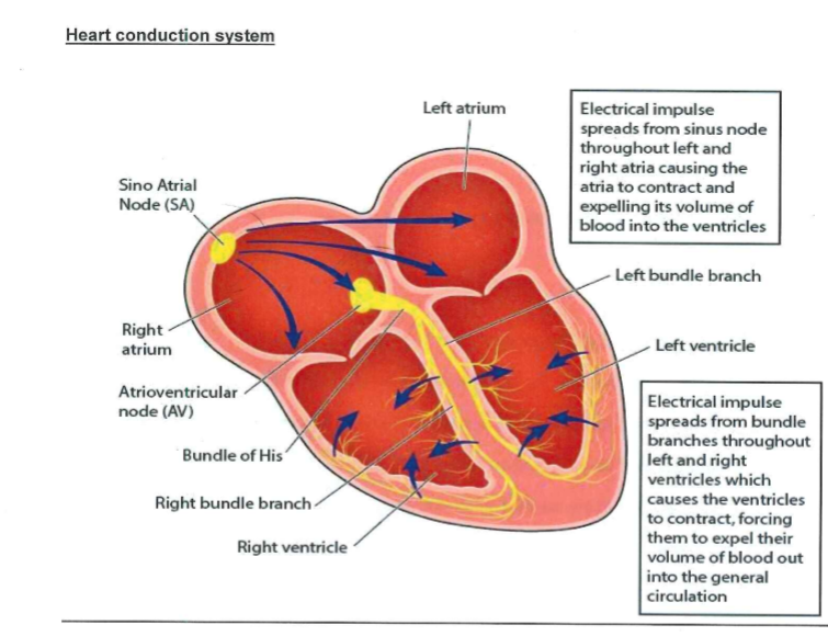
Impulses start at the sinoatrial (SA) node, travel through the atria, causing them to contract (squeeze).  Next the signal travels to the atrioventricular (AV) node, through the bundle of His, down the bundle branches and through the Purkinje fibres, causing the ventricles to contract.

A normal 'sinus' ECG trace


P wave = atria contracting

QRC complex = ventricles contracting

T wave = ventricles relax


What can a 12 lead ECG show us?

  • A heartbeat that is: too fast or too slow, an irregular heartbeat
  • heart muscle that is too thick or parts of the heart that are too big
  • lack of blood flow to the heart muscle
  • inflammation of the sac that surrounds the heart


The ECG leads

You will notice that there are only 10 leads that get attached to the patient, therefore how does it get called a 12 lead ECG?


There are:

six chest leads: V1 V2 V3 V4 V5 V6

six limb leads: aVR aVL aVF I II III (the black ground lead does not record, it acts as a filter to background noise)


Lead I is generated from impulses between left arm and right arm

Lead II is generated from impulses between left leg and right arm

Lead III is generated from impulses between left leg and left arm

Placement of electrodes

Limb leads

  • aVR = right arm (red)
  • aVL = left arm (lemon)
  • aVF = left leg (green)
  • ground electrode = right leg (black)


Chest leads

V1 = 4th intercostal space, right sternal border (red)

V2 = 4th intercostal space, left sternal border (lemon)

V3 = 5th intercostal space, midway between V2 and V4 (green)

V4 = 5th intercostal space, midclavicular line (brown)

V5 = 5th intercostal space, anterior axillary line (black)

V6 = 5th intercostal space, midaxillary line (purple)

How to perform 12 lead ECG

  • Perform hand hygiene before approaching patient
  • Introduce yourself and state why you are there
  • Ensure you have the correct patient - ask them their name and DOB, and/or check wrist band
  • Obtain consent to perform ECG or ensure Adults with Incapacity Act form has been signed
  • Explain procedure to patient (as applicable)
  • Enter patient details into ECG machine
  • Skin needs to be dry, hairless and oil free to allow proper electrode contact.  You may need to shave the patient.  Use an alcohol wipe or abrasive tape to help electrodes stick to skin
  • Position patient sitting at 30 degree angle if possible with arms relaxed by sides and legs uncrossed
  • Place electrodes on patient as above and attach leads
  • Ask patient to keep still and not talk during recording in order to reduce artefact (as applicable)
  • Remove leads and electrodes, ensuring patient is covered up and comfortable.
  • Wash hands.
  • Place ECG in patient's red folder and inform medical/nursing staff
  • Clean ECG machine and leave on charge
  • Do not place electrodes on skin over bones, incisions, irritated skin and body parts where there is a lot of muscle movement

Editorial Information

Last reviewed: 31/01/2021

Next review date: 31/03/2024

Author(s): Williams A.

Author email(s): angela.williams2@borders.scot.nhs.uk.

Reviewer name(s): Williams A.

Related guidelines